Quick answer
This microscope skills worksheet practices labeling microscope parts, calculating total magnification, preparing specimens, focusing safely, and interpreting observations.
Study next
Practice microscope safety, magnification calculations, focusing technique, field of view, and specimen preparation for clear observation.
Read each problem carefully. Show calculations when needed and answer in complete sentences.
Using compound microscopes, calculating magnification, and preparing clear specimens
Science - Grade 9-12
- 1
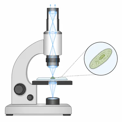
A compound light microscope has a 10x eyepiece and a 40x objective lens. What is the total magnification?
- 2
A student views a specimen first under 100x total magnification and then under 400x total magnification. Describe two changes the student should notice in the image.
- 3
A microscope has a 10x eyepiece. The available objective lenses are 4x, 10x, and 40x. List the total magnifications from lowest to highest.
- 4
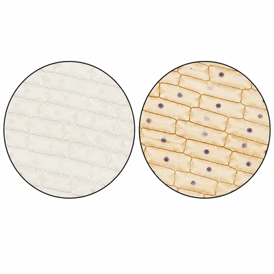
A prepared slide label says the onion cells are stained with iodine. Explain why a stain is often used when viewing cells under a light microscope.
- 5
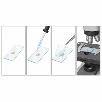
Put these wet mount steps in the correct order: place the specimen on the slide, add a drop of water, lower the coverslip at an angle, place the slide on the stage.
- 6
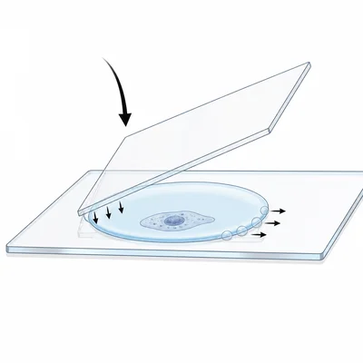
Why should a coverslip be lowered slowly at an angle when making a wet mount?
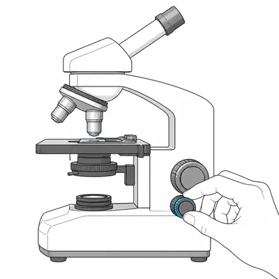
- 7
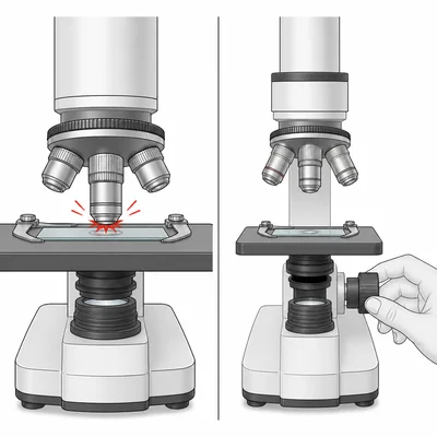
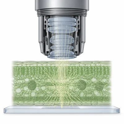
A student begins focusing a slide using the 40x high-power objective and the coarse adjustment knob. Explain the problem with this technique and describe the safer method.
- 8
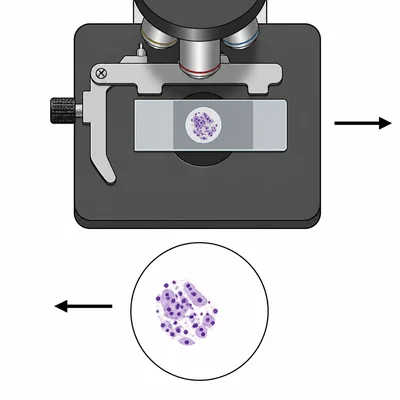
When a specimen moves left in the field of view, which direction did the student move the slide on the stage?
- 9
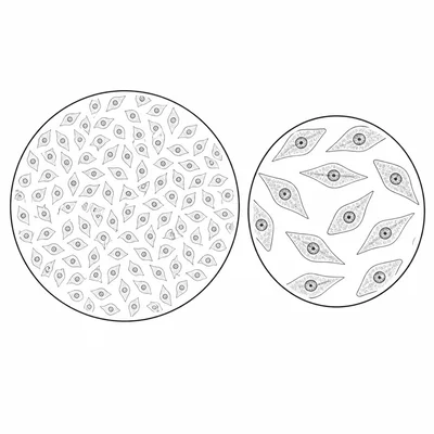
A student estimates that the field of view is 4.0 millimeters wide at 40x total magnification. About how wide is the field of view at 400x total magnification?
- 10
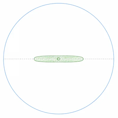
A cell appears to take up about one-fourth of a 0.8 millimeter field of view. Estimate the cell's length in millimeters and micrometers.
- 11
Explain why a very thick specimen may be difficult to observe clearly with a compound light microscope.
- 12
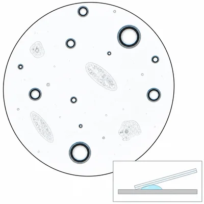
A student sees many dark circles with bright edges in a wet mount. The circles move when the coverslip is gently tapped. What are these circles most likely to be, and how could the student reduce them next time?
- 13
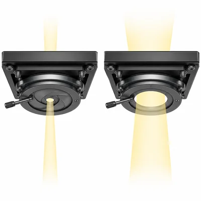
What is the purpose of the diaphragm or iris on a compound light microscope?
- 14
A student switches from the 10x objective to the 40x objective. The image is almost in focus but slightly blurry. Which adjustment knob should the student use, and why?
- 15
Design a brief procedure for preparing and viewing a wet mount of cheek cells. Include specimen collection, slide preparation, staining, and focusing.