Click image to open full size
Biology
Grade 10-12
Microscopy & Cell Imaging Techniques Reference Cheat Sheet
A printable reference covering microscope parts, magnification, resolution, stains, electron microscopy, fluorescence imaging, and scale bars for grades 10-12.
Related Worksheets
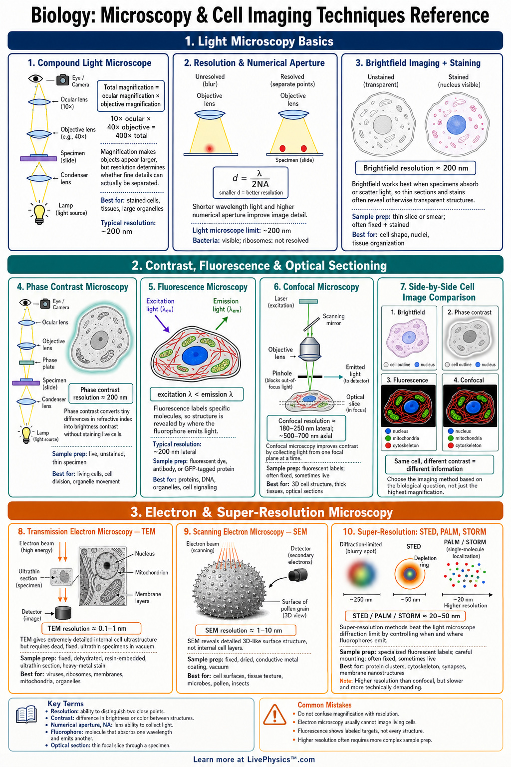
Microscopy and cell imaging techniques help students observe structures that are too small to see with the unaided eye. This cheat sheet summarizes the tools, measurements, and imaging methods used to study cells and tissues. Students need it to compare microscope types, calculate magnification, and interpret cell images accurately. It is especially useful for biology labs, microscopy practicals, and exam review.
Key Facts
- Total magnification = eyepiece magnification x objective magnification.
- Resolution is the ability to distinguish two close points as separate, and higher resolution shows finer detail.
- Field of view usually decreases as magnification increases, so less of the specimen is visible at high power.
- Actual size = image size / magnification when image size and actual size use the same units.
- Light microscopes can view living cells, but electron microscopes usually require dead, specially prepared specimens.
- Transmission electron microscopes show internal cell structures, while scanning electron microscopes show surface detail.
- Stains and dyes increase contrast by binding to specific cell parts, such as nuclei, membranes, or cell walls.
- A scale bar on a micrograph shows the real length represented in the image and is often more reliable than stated magnification.
Vocabulary
- Magnification
- Magnification is how many times larger an image appears compared with the specimen's actual size.
- Resolution
- Resolution is the ability of a microscope or imaging system to show two nearby points as separate.
- Field of View
- Field of view is the visible area seen through the microscope at a given magnification.
- Contrast
- Contrast is the difference in brightness or color that helps cell structures stand out from the background.
- Fluorescence Microscopy
- Fluorescence microscopy uses fluorescent molecules that absorb light at one wavelength and emit light at another.
- Micrograph
- A micrograph is a photograph or digital image taken through a microscope.
Common Mistakes to Avoid
- Using only the objective lens power as total magnification is wrong because total magnification also includes the eyepiece lens.
- Confusing magnification with resolution is wrong because a larger image is not always a clearer or more detailed image.
- Forgetting to convert units before using actual size = image size / magnification is wrong because mixed units give an incorrect answer.
- Using the coarse focus knob on high power is wrong because it can crash the objective lens into the slide and damage the specimen.
- Assuming all microscope images show natural colors is wrong because stains, dyes, and digital processing often create artificial color.
Practice Questions
- 1 A microscope has a 10x eyepiece and a 40x objective lens. What is the total magnification?
- 2 A cell image measures 60 mm on a printout and the magnification is 3000x. What is the actual cell size in mm?
- 3 A scale bar labeled 10 micrometers measures 25 mm on a printed micrograph. If a mitochondrion measures 12.5 mm on the same image, what is its actual length?
- 4 Why might a biologist choose fluorescence microscopy instead of a standard brightfield light microscope when studying protein location inside cells?