Practice the physics behind three major medical imaging methods, X-rays, ultrasound, and MRI. Explore wave behavior, absorption, reflection, frequency, wavelength, safety, and image contrast.
Read each problem carefully. Show calculations when needed, include units, and explain your reasoning in complete sentences.
Comparing how electromagnetic waves, sound waves, and magnetic fields create medical images
Physics - Grade 9-12
- 1
An X-ray photon has a frequency of 3.0 x 10^18 Hz. Calculate its wavelength in meters using c = 3.0 x 10^8 m/s.
- 2
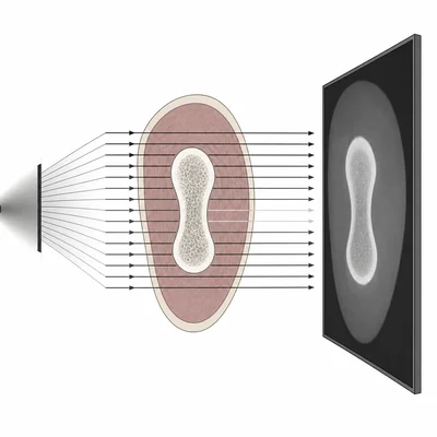
Explain why bones appear lighter than soft tissue on a typical X-ray image.
- 3
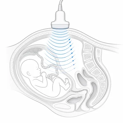
A technician wants to image a fetus during pregnancy. Which imaging method is usually preferred: X-ray, ultrasound, or MRI? Explain why.
- 4
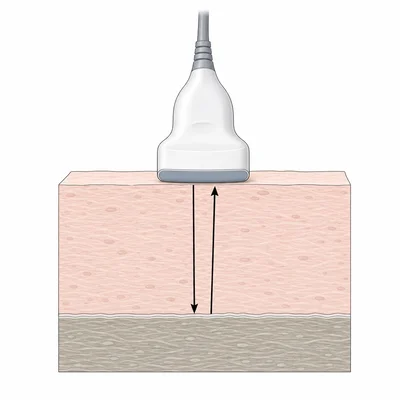
Ultrasound travels through soft tissue at about 1540 m/s. If an ultrasound pulse returns from a boundary after 80 microseconds, how deep is the boundary? Remember that the pulse travels to the boundary and back.
- 5
Describe one way an MRI is different from an X-ray in the type of physics it uses to make an image.
- 6
A medical ultrasound probe uses a frequency of 5.0 MHz. If sound travels in tissue at 1540 m/s, what is the wavelength in tissue?
- 7
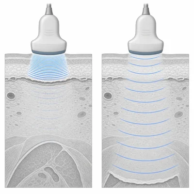
Higher-frequency ultrasound can produce more detailed images but does not travel as deeply into the body. Explain this tradeoff.
- 8
Match each imaging method to the main type of wave or field it uses: X-ray, ultrasound, and MRI.
- 9
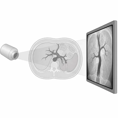
A radiologist uses contrast material during an X-ray or CT scan. Explain what contrast material does in terms of image formation.
- 10
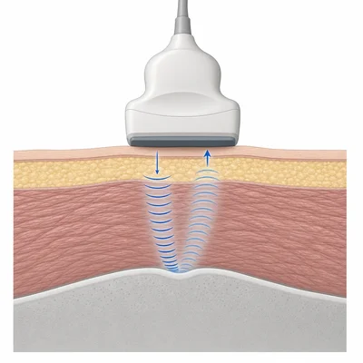
In ultrasound imaging, a strong echo occurs at a boundary between two materials with very different acoustic properties. Give an example of a boundary that would create a strong echo and explain why.
- 11
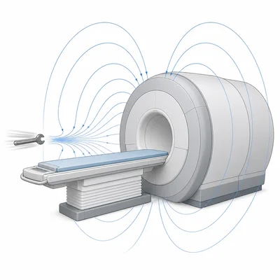
Why must patients remove metal objects before an MRI scan?
- 12
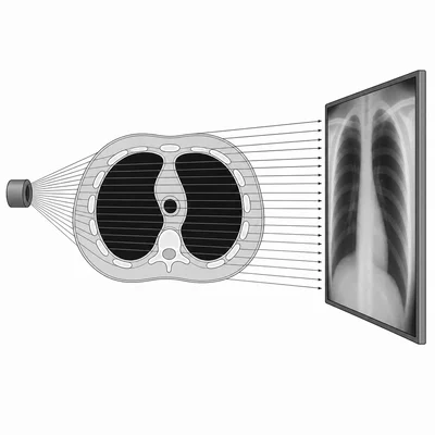
An X-ray image shows high contrast between bone and lung tissue. Explain why lung tissue often appears darker than many other body tissues.
- 13
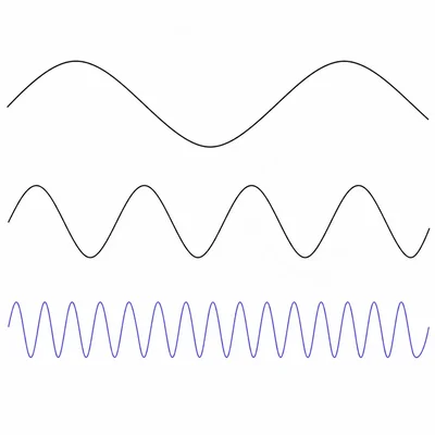
Rank X-rays, visible light, and radio waves from lowest photon energy to highest photon energy. Explain using frequency.
- 14
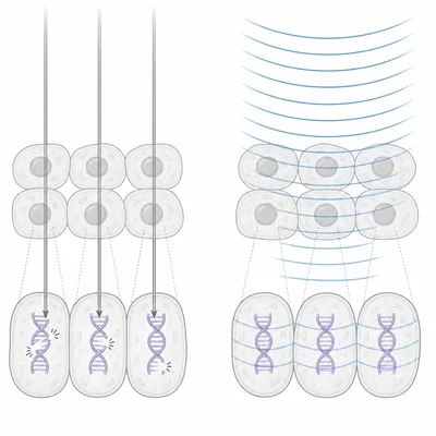
A patient asks why repeated X-ray scans are limited when possible, but ultrasound exams do not have the same radiation concern. Write a physics-based explanation.
- 15
Compare the best use of X-ray, ultrasound, and MRI for imaging the body. Give one example of a situation where each method is useful.