Understanding 3D Human Anatomy Explorer
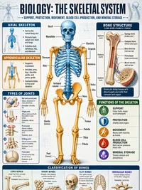
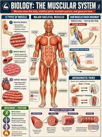
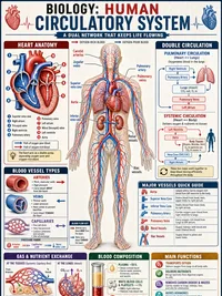
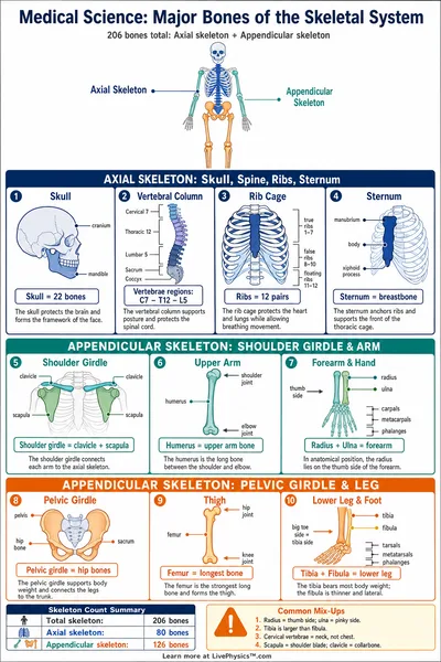
This 3D human anatomy explorer shows the body in six layers that you can switch on and off. The layers are the skin, the skeleton, the muscles, the organs, the arteries and the veins.
Drag to turn the body, zoom in, and click any bone, muscle, organ or blood vessel to see its name. More than 1,500 structures carry their anatomical name, and the main ones have an everyday name too, so the femur is also listed as the thigh bone.
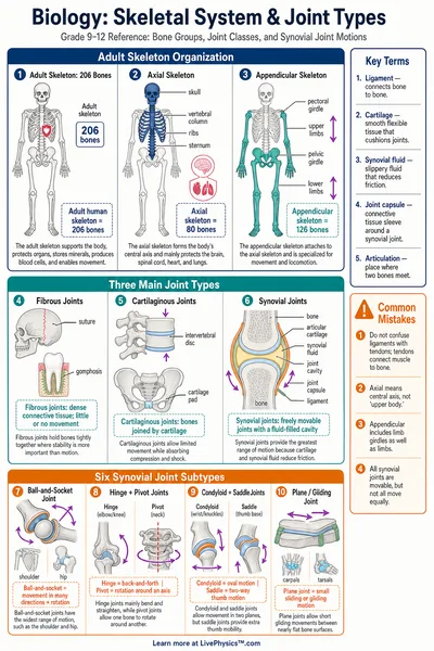
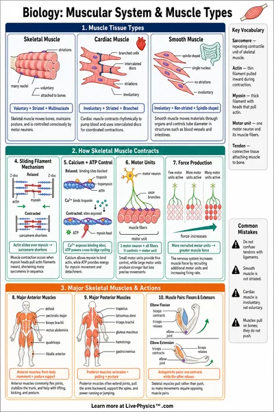
Layers help because the body is packed in depth. The skin covers everything, muscles cover most of the skeleton, and the ribs form a cage around the heart and the lungs. Switching a layer off shows what lies underneath it.
You can hide a structure, show it alone, or fade the rest of the body to see where it sits. Fading works well for deep structures such as the kidneys, which lie at the back of the abdomen behind the intestines.
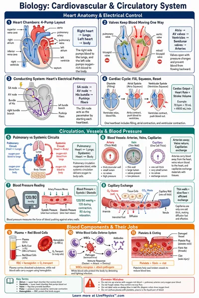
Arteries are drawn in red and veins in blue. Arteries carry blood away from the heart and veins carry it back. The colors are a convention from anatomy diagrams. Blood in the veins is dark red, not blue.
Left and right always describe the body's own sides. When the model faces you, its left arm is on the right side of your screen.
Quiz mode names a structure and asks you to find it on the model. You can answer with a click, with a tap, or with the arrow keys and Enter. Each round has ten questions drawn from the main structures that can be seen from outside in the layers you switched on, so you can practice one body system at a time. The skin layer switches off while a quiz runs.
Any view can be copied as a link that opens with the same layers, the same selection and the same camera angle. A teacher can prepare a view and send it to a class.
The models come from BodyParts3D, an open data set that models an adult male body. This version does not include the nerves or the hair. Real bodies also vary in size and in the way blood vessels branch, so treat the model as a typical example.