Understanding Nephron Filtration & Osmoregulation Lab
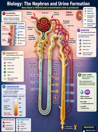
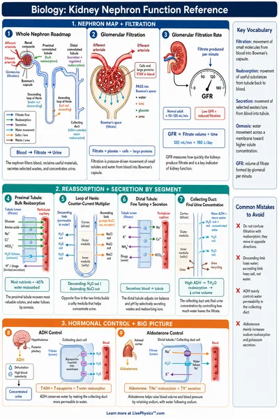
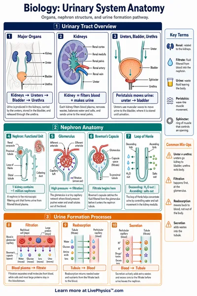
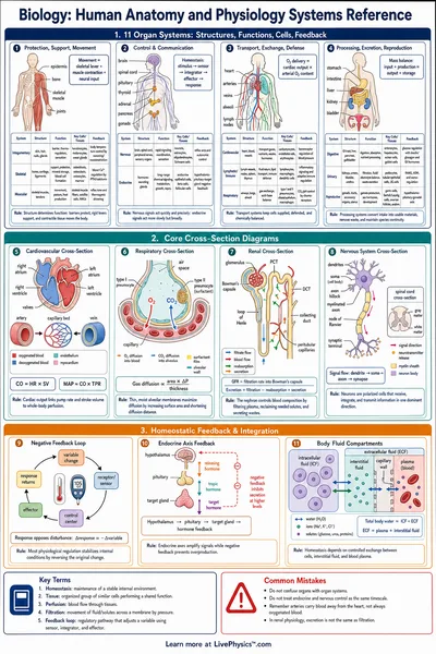
Filtration begins because pressure inside tiny glomerular capillaries pushes water and small dissolved substances across a selective barrier. Blood cells and most proteins stay in the bloodstream because they are too large or have electrical properties that the barrier resists.
The pressure depends on the balance between blood entering and leaving the glomerulus, not on a blood pressure reading alone. This is why changing the width of nearby blood vessels can change filtration while overall blood pressure appears stable.
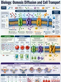
The early tubule does much more than carry fluid onward. Its cells use energy to move sodium out of the filtrate, and water follows those dissolved particles by osmosis. Useful substances such as amino acids, bicarbonate, and much of the filtered glucose are taken back into the blood here.
This recovery prevents the body from wasting valuable nutrients and helps keep blood volume within a safe range. Some medicines act on tubular transport, which explains why certain drugs increase urine production.
The loop of Henle builds a salt concentration gradient in the kidney tissue. Its descending side allows water to leave more easily, while its ascending side moves salts outward but does not allow water to follow. This arrangement makes the deep kidney tissue unusually concentrated.
When the body needs to save water, fluid later passing through the collecting duct can release water into this concentrated area. The result is a smaller volume of darker, more concentrated urine during dehydration.
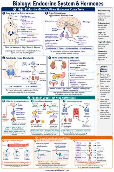
ADH changes how freely water can cross cells in the collecting duct. High ADH places more water channels in these cell membranes, so water returns to the blood instead of remaining in urine. Low ADH leaves fewer channels available, producing a larger volume of dilute urine.
Alcohol can reduce ADH release, which is one reason people often urinate more after drinking it. This response can worsen dehydration because fluid is lost when the body should be conserving it.
Glucose reabsorption has a transport limit because each carrier protein can work only so fast. Once blood glucose rises beyond that limit, glucose stays in the filtrate and appears in urine. The extra glucose holds water in the tubule, so urine volume increases and thirst may follow.
This pattern is important in untreated diabetes mellitus. In the lab, pay attention to which variable changes first and which output follows, since urine volume, sodium loss, and concentration do not always move in the same direction.