
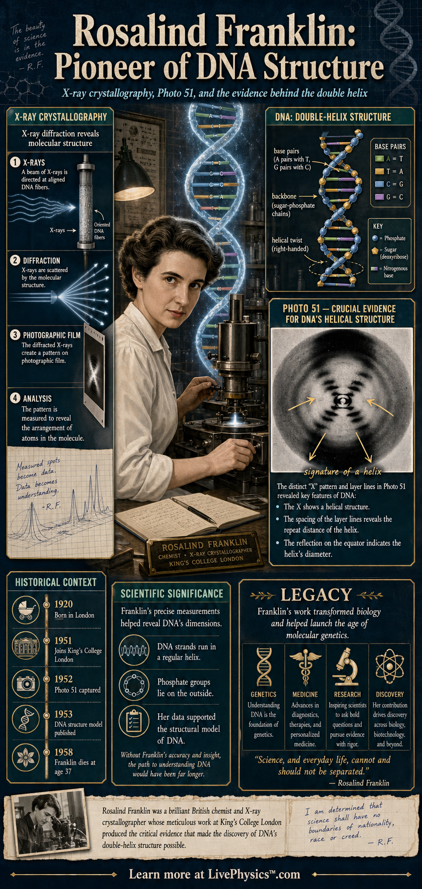
Rosalind Franklin was a British chemist whose careful X-ray crystallography helped reveal the structure of DNA. Her famous diffraction image, known as Photo 51, gave strong evidence that DNA has a helical shape. Franklin also made important discoveries about coal, graphite, and viruses, showing how chemistry connects atomic structure to real materials.
Her work matters because modern biology, genetics, and molecular medicine all depend on knowing how molecules are arranged in three dimensions.
X-ray crystallography works by sending X-rays through an ordered sample and recording the pattern made when the waves scatter. The spacing and angles in the pattern can be used to infer distances between atoms or repeating features in a molecule. At King's College London, Franklin and her student Raymond Gosling produced high quality DNA diffraction images that supported key measurements used in building the double helix model.
Although her contribution to DNA structure was understated during her lifetime, her experimental skill is now recognized as essential to one of the greatest scientific breakthroughs of the twentieth century.
Understanding Rosalind Franklin, Pioneer of X-Ray Crystallography
A diffraction photograph is not a direct picture of atoms. It is a record of where many scattered X-ray waves reinforce or cancel each other. Each dark spot carries information about a repeating arrangement inside the sample.
The position of spots gives distances. Their brightness gives clues about how matter is distributed.
Scientists must turn this indirect evidence into a structure, often by comparing many possible models with the measured pattern. This makes crystallography a careful process of measurement, calculation, and checking rather than a single photograph that gives an instant answer.
DNA presented a special challenge because its shape changed with water content. Franklin distinguished a drier A form from a wetter B form. The B form produced the especially clear helical pattern associated with Photo 51.
From the spacing of features in the diffraction pattern, she could determine the regular distance between stacked bases and the larger repeat distance along the molecule. She found that the phosphate groups were likely on the outside, where they could interact with water.
These constraints ruled out many incorrect arrangements. A successful model had to fit every important measurement, not merely look like a plausible helix.
This lesson matters across chemistry. A material behaves differently when its particles are arranged differently. In coal, tiny pores and ordered carbon regions affect how gases move through the material and how it burns.
In graphite, layers of carbon atoms can slide over one another, which helps explain its use in pencil leads and lubricants. Virus research uses the same structural idea.
A virus can only infect a cell if its proteins fit together in precise ways. Knowing their arrangement helps researchers understand how a virus is built, how it carries genetic material, and where a treatment might interfere with its function.
Students often meet these ideas when learning about waves, atomic structure, polymers, and biological molecules. The important habit is to separate evidence from interpretation. An X shaped diffraction pattern supports a helix, but it does not by itself supply every detail of a molecular model.
Measurements have uncertainty. Samples can contain water, impurities, or mixed forms that complicate a result. Good scientists record conditions, repeat observations, and test whether different pieces of evidence agree.
Franklin's work shows why patient experimental work is essential. Clear data can place strict limits on what nature allows, even before the final structure is fully understood.
Key Facts
- Rosalind Franklin lived from 1920 to 1958 and was trained as a physical chemist.
- Photo 51 was an X-ray diffraction image of DNA that showed an X shaped pattern, a signature of a helix.
- Bragg's law connects diffraction angle to spacing, nλ = 2d sin θ.
- Franklin's DNA work at King's College London helped identify key dimensions of the double helix.
- She also studied the microstructure of coal and graphite, linking material properties to atomic arrangement.
- Franklin later investigated viruses such as tobacco mosaic virus and polio using X-ray methods.
Vocabulary
- X-ray crystallography
- A method for determining molecular structure by analyzing how X-rays scatter from an ordered sample.
- Diffraction pattern
- A pattern of spots or bands formed when waves scatter and interfere after passing through or reflecting from a structure.
- Photo 51
- A famous X-ray diffraction image of DNA taken by Raymond Gosling under Rosalind Franklin's supervision.
- Double helix
- The twisted ladder shape of DNA made from two strands wrapped around the same axis.
- Bragg's law
- A relationship that uses X-ray wavelength and diffraction angle to calculate spacing between repeating planes in a structure.
Common Mistakes to Avoid
- Thinking Photo 51 was a normal photograph, which is wrong because it is a diffraction pattern that must be mathematically interpreted.
- Assuming Franklin only worked on DNA, which is wrong because she also made major contributions to coal, graphite, tobacco mosaic virus, and polio virus research.
- Treating an X shaped diffraction pattern as direct proof of every detail of DNA structure, which is wrong because it mainly indicates helical symmetry and must be combined with measurements and chemical reasoning.
- Forgetting the units in Bragg's law calculations, which is wrong because wavelength and spacing must be in the same length units for d to be meaningful.
Practice Questions
- 1 Using Bragg's law nλ = 2d sin θ, find d for first order diffraction when λ = 0.154 nm and θ = 15.0 degrees.
- 2 Photo 51 was produced in 1952. How old was Rosalind Franklin in 1952 if she was born in 1920?
- 3 Explain why an X shaped diffraction pattern supports the idea that DNA is helical rather than a straight, flat molecule.