Muscle Contraction Sliding-Filament Lab
Calcium exposes the actin binding sites, the sarcomere length sets how well actin and myosin overlap, and ATP powers the cross-bridge cycle. Change all three and see how many cross-bridges form and how much force the muscle fibre produces.
Guided ExperimentCalcium and the activation curve
If you raise the calcium concentration, how does the relative force change, and what shape is the curve?
Write your hypothesis in the Lab Report panel, then click Next.
Controls
Sarcomere
2.10 µmContraction results
Relative force
50% of max
Calcium activation
50%
Length-tension
100%
Active cross-bridges
12of 24
Shortening velocity
50% of max
ATP state
Cycling
Length-tension relationship
Calcium activation (Hill curve)
What this means
- At the optimal sarcomere length the actin and myosin overlap is ideal, giving maximum force.
- Stretching the sarcomere too far removes the overlap, so force drops to zero even with plenty of calcium.
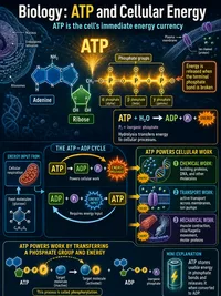
- Without ATP the cross-bridges lock in rigor and cannot cycle, so the muscle cannot produce or release force normally.
The cross-bridge cycle
The cocked myosin head binds an exposed site on the actin filament, forming a cross-bridge.
The head pivots and pulls the thin filament toward the centre, releasing ADP and phosphate.
A new ATP molecule binds the myosin head, which lets the head release from actin.
ATP is hydrolysed to ADP and phosphate, re-cocking the head so it is ready to bind again.
Steps 3 and 4 both need ATP. Without it the head stays bound to actin and the muscle locks in rigor.
Data Table
(0 rows)| # | Calcium(µM) | ATP(%) | Sarcomere(µm) | Activation(%) | Length-tension(%) | Force(%) |
|---|
Reference Guide
The Sliding-Filament Theory
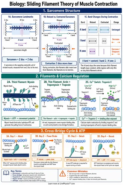
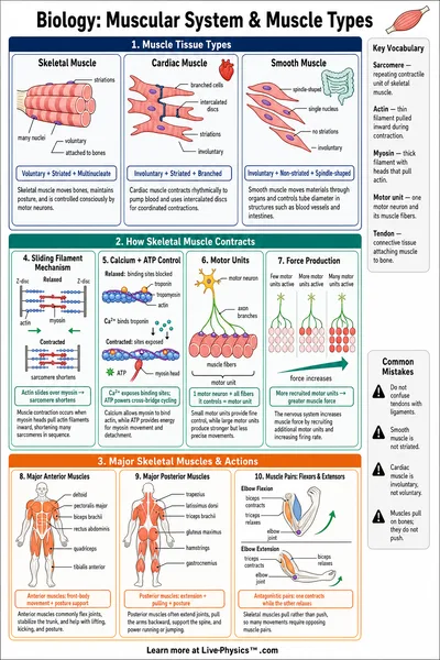
A muscle fibre is made of repeating units called sarcomeres. Each sarcomere is bounded by two Z-discs and contains thin and thick filaments that slide past one another.
- Actin. The thin filament anchored at the Z-discs.
- Myosin. The thick filament in the centre, with heads that reach to actin.
- Z-discs. The boundaries of each sarcomere.
- Contraction. Myosin heads pull actin inward, so the Z-discs move closer and the sarcomere shortens.
The filaments themselves do not shorten. They slide, which is why this is called the sliding-filament model.
Calcium and Turning Contraction On
At rest, tropomyosin covers the actin binding sites so myosin cannot attach. Calcium is the switch that uncovers them.
- Calcium binds troponin. A nerve signal releases calcium from the sarcoplasmic reticulum.
- Tropomyosin shifts. Troponin moves tropomyosin off the binding sites.
- Steep response. Because several calcium ions are involved, the activation curve is steep and switch-like.
In this lab the calcium activation follows a Hill curve that is about half maximal near 1 micromolar calcium.
The Cross-Bridge Cycle and ATP
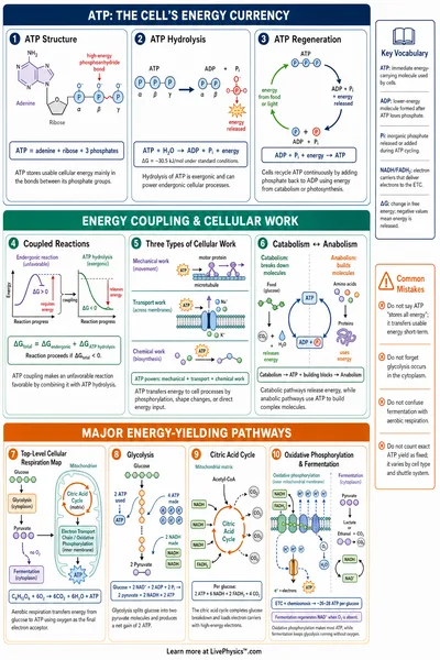
Each myosin head attaches to actin, pulls, detaches, and re-cocks. ATP is needed twice in every cycle.
- Attach. The cocked head binds an exposed actin site.
- Power stroke. The head pivots and pulls the thin filament.
- Detach. A new ATP binds the head so it releases from actin.
- Re-cock. ATP is hydrolysed, recharging the head for another cycle.
Without ATP the head cannot detach, so the muscle locks in rigor. This is what produces rigor mortis after death.
The Length-Tension Relationship
The force a sarcomere can produce depends on how well its thin and thick filaments overlap, which depends on its length.
- Optimal length. Near 2.0 to 2.25 micrometres the overlap is ideal and force is maximal.
- Too short. Below about 2.0 micrometres the filaments collide and force falls toward zero near 1.27 micrometres.
- Too long. Above the plateau the overlap shrinks, reaching zero force near 3.65 micrometres.
Even with full calcium and ATP, a sarcomere stretched too far cannot form cross-bridges, so it produces almost no force.